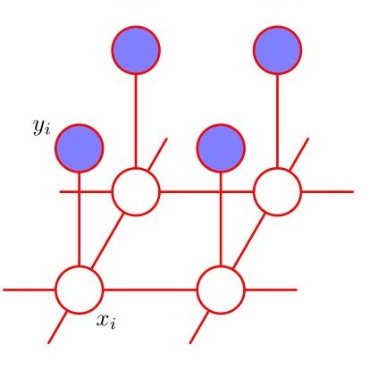
Image segmentation is the process of partitioning the image into significant regions easier to analyze. Nowadays, segmentation has become a necessity in many practical medical imaging methods as locating tumors and diseases. Hidden Markov Random Field model is one of several techniques used in image segmentation. It provides an elegant way to model the segmentation process. This modeling leads to the minimization of an objective function. Conjugate Gradient algorithm (CG) is one of the best known optimization techniques. This paper proposes the use of the Conjugate Gradient algorithm (CG) for image segmentation, based on the Hidden Markov Random Field. Since derivatives are not available for this expression, finite differences are used in the CG algorithm to approximate the first derivative. The approach is evaluated using a number of publicly available images, where ground truth is known. The Dice Coefficient is used as an objective criterion to measure the quality of segmentation. The results show that the proposed CG approach compares favorably with other variants of Hidden Markov Random Field segmentation algorithms.
相關內容
We propose a novel technique to incorporate attention within convolutional neural networks using feature maps generated by a separate convolutional autoencoder. Our attention architecture is well suited for incorporation with deep convolutional networks. We evaluate our model on benchmark segmentation datasets in skin cancer segmentation and lung lesion segmentation. Results show highly competitive performance when compared with U-Net and it's residual variant.
An important step in early brain development study is to perform automatic segmentation of infant brain magnetic resonance (MR) images into cerebrospinal fluid (CSF), gray matter (GM) and white matter (WM) regions. This task is especially challenging in the isointense stage (approximately 6-8 months of age) when GM and WM exhibit similar levels of intensities in MR images. Deep learning has shown its great promise in various image segmentation tasks. However, existing models do not have an efficient and effective way to aggregate global information. They also suffer from information loss during up-sampling operations. In this work, we address these problems by proposing a global aggregation block, which can be flexibly used for global information fusion. We build a novel model based on 3D U-Net to make fast and accurate voxel-wise dense prediction. We perform thorough experiments, and results indicate that our model outperforms previous best models significantly on 3D multimodality isointense infant brain MR image segmentation.
In two-phase image segmentation, convex relaxation has allowed global minimisers to be computed for a variety of data fitting terms. Many efficient approaches exist to compute a solution quickly. However, we consider whether the nature of the data fitting in this formulation allows for reasonable assumptions to be made about the solution that can improve the computational performance further. In particular, we employ a well known dual formulation of this problem and solve the corresponding equations in a restricted domain. We present experimental results that explore the dependence of the solution on this restriction and quantify imrovements in the computational performance. This approach can be extended to analogous methods simply and could provide an efficient alternative for problems of this type.
The piecewise constant Mumford-Shah (PCMS) model and the Rudin-Osher-Fatemi (ROF) model are two of the most famous variational models in image segmentation and image restoration, respectively. They have ubiquitous applications in image processing. In this paper, we explore the linkage between these two important models. We prove that for two-phase segmentation problem the optimal solution of the PCMS model can be obtained by thresholding the minimizer of the ROF model. This linkage is still valid for multiphase segmentation under mild assumptions. Thus it opens a new segmentation paradigm: image segmentation can be done via image restoration plus thresholding. This new paradigm, which circumvents the innate non-convex property of the PCMS model, therefore improves the segmentation performance in both efficiency (much faster than state-of-the-art methods based on PCMS model, particularly when the phase number is high) and effectiveness (producing segmentation results with better quality) due to the flexibility of the ROF model in tackling degraded images, such as noisy images, blurry images or images with information loss. As a by-product of the new paradigm, we derive a novel segmentation method, coined thresholded-ROF (T-ROF) method, to illustrate the virtue of manipulating image segmentation through image restoration techniques. The convergence of the T-ROF method under certain conditions is proved, and elaborate experimental results and comparisons are presented.
The Conditional Random Field as a Recurrent Neural Network layer is a recently proposed algorithm meant to be placed on top of an existing Fully-Convolutional Neural Network to improve the quality of semantic segmentation. In this paper, we test whether this algorithm, which was shown to improve semantic segmentation for 2D RGB images, is able to improve segmentation quality for 3D multi-modal medical images. We developed an implementation of the algorithm which works for any number of spatial dimensions, input/output image channels, and reference image channels. As far as we know this is the first publicly available implementation of this sort. We tested the algorithm with two distinct 3D medical imaging datasets, we concluded that the performance differences observed were not statistically significant. Finally, in the discussion section of the paper, we go into the reasons as to why this technique transfers poorly from natural images to medical images.
Over the past decades, state-of-the-art medical image segmentation has heavily rested on signal processing paradigms, most notably registration-based label propagation and pair-wise patch comparison, which are generally slow despite a high segmentation accuracy. In recent years, deep learning has revolutionalized computer vision with many practices outperforming prior art, in particular the convolutional neural network (CNN) studies on image classification. Deep CNN has also started being applied to medical image segmentation lately, but generally involves long training and demanding memory requirements, achieving limited success. We propose a patch-based deep learning framework based on a revisit to the classic neural network model with substantial modernization, including the use of Rectified Linear Unit (ReLU) activation, dropout layers, 2.5D tri-planar patch multi-pathway settings. In a test application to hippocampus segmentation using 100 brain MR images from the ADNI database, our approach significantly outperformed prior art in terms of both segmentation accuracy and speed: scoring a median Dice score up to 90.98% on a near real-time performance (<1s).
One of the most common tasks in medical imaging is semantic segmentation. Achieving this segmentation automatically has been an active area of research, but the task has been proven very challenging due to the large variation of anatomy across different patients. However, recent advances in deep learning have made it possible to significantly improve the performance of image recognition and semantic segmentation methods in the field of computer vision. Due to the data driven approaches of hierarchical feature learning in deep learning frameworks, these advances can be translated to medical images without much difficulty. Several variations of deep convolutional neural networks have been successfully applied to medical images. Especially fully convolutional architectures have been proven efficient for segmentation of 3D medical images. In this article, we describe how to build a 3D fully convolutional network (FCN) that can process 3D images in order to produce automatic semantic segmentations. The model is trained and evaluated on a clinical computed tomography (CT) dataset and shows state-of-the-art performance in multi-organ segmentation.
Image segmentation is considered to be one of the critical tasks in hyperspectral remote sensing image processing. Recently, convolutional neural network (CNN) has established itself as a powerful model in segmentation and classification by demonstrating excellent performances. The use of a graphical model such as a conditional random field (CRF) contributes further in capturing contextual information and thus improving the segmentation performance. In this paper, we propose a method to segment hyperspectral images by considering both spectral and spatial information via a combined framework consisting of CNN and CRF. We use multiple spectral cubes to learn deep features using CNN, and then formulate deep CRF with CNN-based unary and pairwise potential functions to effectively extract the semantic correlations between patches consisting of three-dimensional data cubes. Effective piecewise training is applied in order to avoid the computationally expensive iterative CRF inference. Furthermore, we introduce a deep deconvolution network that improves the segmentation masks. We also introduce a new dataset and experimented our proposed method on it along with several widely adopted benchmark datasets to evaluate the effectiveness of our method. By comparing our results with those from several state-of-the-art models, we show the promising potential of our method.
Precise 3D segmentation of infant brain tissues is an essential step towards comprehensive volumetric studies and quantitative analysis of early brain developement. However, computing such segmentations is very challenging, especially for 6-month infant brain, due to the poor image quality, among other difficulties inherent to infant brain MRI, e.g., the isointense contrast between white and gray matter and the severe partial volume effect due to small brain sizes. This study investigates the problem with an ensemble of semi-dense fully convolutional neural networks (CNNs), which employs T1-weighted and T2-weighted MR images as input. We demonstrate that the ensemble agreement is highly correlated with the segmentation errors. Therefore, our method provides measures that can guide local user corrections. To the best of our knowledge, this work is the first ensemble of 3D CNNs for suggesting annotations within images. Furthermore, inspired by the very recent success of dense networks, we propose a novel architecture, SemiDenseNet, which connects all convolutional layers directly to the end of the network. Our architecture allows the efficient propagation of gradients during training, while limiting the number of parameters, requiring one order of magnitude less parameters than popular medical image segmentation networks such as 3D U-Net. Another contribution of our work is the study of the impact that early or late fusions of multiple image modalities might have on the performances of deep architectures. We report evaluations of our method on the public data of the MICCAI iSEG-2017 Challenge on 6-month infant brain MRI segmentation, and show very competitive results among 21 teams, ranking first or second in most metrics.


