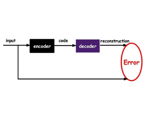
Unsupervised anomaly detection in medical images such as chest radiographs is stepping into the spotlight as it mitigates the scarcity of the labor-intensive and costly expert annotation of anomaly data. However, nearly all existing methods are formulated as a one-class classification trained only on representations from the normal class and discard a potentially significant portion of the unlabeled data. This paper focuses on a more practical setting, dual distribution anomaly detection for chest X-rays, using the entire training data, including both normal and unlabeled images. Inspired by a modern self-supervised vision transformer model trained using partial image inputs to reconstruct missing image regions -- we propose AMAE, a two-stage algorithm for adaptation of the pre-trained masked autoencoder (MAE). Starting from MAE initialization, AMAE first creates synthetic anomalies from only normal training images and trains a lightweight classifier on frozen transformer features. Subsequently, we propose an adaptation strategy to leverage unlabeled images containing anomalies. The adaptation scheme is accomplished by assigning pseudo-labels to unlabeled images and using two separate MAE based modules to model the normative and anomalous distributions of pseudo-labeled images. The effectiveness of the proposed adaptation strategy is evaluated with different anomaly ratios in an unlabeled training set. AMAE leads to consistent performance gains over competing self-supervised and dual distribution anomaly detection methods, setting the new state-of-the-art on three public chest X-ray benchmarks: RSNA, NIH-CXR, and VinDr-CXR.
相關內容
The lack of annotated medical images limits the performance of deep learning models, which usually need large-scale labelled datasets. Few-shot learning techniques can reduce data scarcity issues and enhance medical image analysis, especially with meta-learning. This systematic review gives a comprehensive overview of few-shot learning in medical imaging. We searched the literature systematically and selected 80 relevant articles published from 2018 to 2023. We clustered the articles based on medical outcomes, such as tumour segmentation, disease classification, and image registration; anatomical structure investigated (i.e. heart, lung, etc.); and the meta-learning method used. For each cluster, we examined the papers' distributions and the results provided by the state-of-the-art. In addition, we identified a generic pipeline shared among all the studies. The review shows that few-shot learning can overcome data scarcity in most outcomes and that meta-learning is a popular choice to perform few-shot learning because it can adapt to new tasks with few labelled samples. In addition, following meta-learning, supervised learning and semi-supervised learning stand out as the predominant techniques employed to tackle few-shot learning challenges in medical imaging and also best performing. Lastly, we observed that the primary application areas predominantly encompass cardiac, pulmonary, and abdominal domains. This systematic review aims to inspire further research to improve medical image analysis and patient care.
Weakly Supervised Semantic Segmentation (WSSS) relying only on image-level supervision is a promising approach to deal with the need for Segmentation networks, especially for generating a large number of pixel-wise masks in a given dataset. However, most state-of-the-art image-level WSSS techniques lack an understanding of the geometric features embedded in the images since the network cannot derive any object boundary information from just image-level labels. We define a boundary here as the line separating an object and its background, or two different objects. To address this drawback, we are proposing our novel ReFit framework, which deploys state-of-the-art class activation maps combined with various post-processing techniques in order to achieve fine-grained higher-accuracy segmentation masks. To achieve this, we investigate a state-of-the-art unsupervised segmentation network that can be used to construct a boundary map, which enables ReFit to predict object locations with sharper boundaries. By applying our method to WSSS predictions, we achieved up to 10% improvement over the current state-of-the-art WSSS methods for medical imaging. The framework is open-source, to ensure that our results are reproducible, and accessible online at //github.com/bharathprabakaran/ReFit.
In the past decade, there has been significant advancement in designing wearable neural interfaces for controlling neurorobotic systems, particularly bionic limbs. These interfaces function by decoding signals captured non-invasively from the skin's surface. Portable high-density surface electromyography (HD-sEMG) modules combined with deep learning decoding have attracted interest by achieving excellent gesture prediction and myoelectric control of prosthetic systems and neurorobots. However, factors like pixel-shape electrode size and unstable skin contact make HD-sEMG susceptible to pixel electrode drops. The sparse electrode-skin disconnections rooted in issues such as low adhesion, sweating, hair blockage, and skin stretch challenge the reliability and scalability of these modules as the perception unit for neurorobotic systems. This paper proposes a novel deep-learning model providing resiliency for HD-sEMG modules, which can be used in the wearable interfaces of neurorobots. The proposed 3D Dilated Efficient CapsNet model trains on an augmented input space to computationally `force' the network to learn channel dropout variations and thus learn robustness to channel dropout. The proposed framework maintained high performance under a sensor dropout reliability study conducted. Results show conventional models' performance significantly degrades with dropout and is recovered using the proposed architecture and the training paradigm.
We present a set of metrics that utilize vision priors to effectively assess the performance of saliency methods on image classification tasks. To understand behavior in deep learning models, many methods provide visual saliency maps emphasizing image regions that most contribute to a model prediction. However, there is limited work on analyzing the reliability of saliency methods in explaining model decisions. We propose the metric COnsistency-SEnsitivity (COSE) that quantifies the equivariant and invariant properties of visual model explanations using simple data augmentations. Through our metrics, we show that although saliency methods are thought to be architecture-independent, most methods could better explain transformer-based models over convolutional-based models. In addition, GradCAM was found to outperform other methods in terms of COSE but was shown to have limitations such as lack of variability for fine-grained datasets. The duality between consistency and sensitivity allow the analysis of saliency methods from different angles. Ultimately, we find that it is important to balance these two metrics for a saliency map to faithfully show model behavior.
We present a new method for automatically classifying medical images that uses weak causal signals in the scene to model how the presence of a feature in one part of the image affects the appearance of another feature in a different part of the image. Our method consists of two components: a convolutional neural network backbone and a causality-factors extractor module. The latter computes weights for the feature maps to enhance each feature map according to its causal influence in the image's scene. We can modify the functioning of the causality module by using two external signals, thus obtaining different variants of our method. We evaluate our method on a public dataset of prostate MRI images for prostate cancer diagnosis, using quantitative experiments, qualitative assessment, and ablation studies. Our results show that our method improves classification performance and produces more robust predictions, focusing on relevant parts of the image. That is especially important in medical imaging, where accurate and reliable classifications are essential for effective diagnosis and treatment planning.
The extraction of structured clinical information from free-text radiology reports in the form of radiology graphs has been demonstrated to be a valuable approach for evaluating the clinical correctness of report-generation methods. However, the direct generation of radiology graphs from chest X-ray (CXR) images has not been attempted. To address this gap, we propose a novel approach called Prior-RadGraphFormer that utilizes a transformer model with prior knowledge in the form of a probabilistic knowledge graph (PKG) to generate radiology graphs directly from CXR images. The PKG models the statistical relationship between radiology entities, including anatomical structures and medical observations. This additional contextual information enhances the accuracy of entity and relation extraction. The generated radiology graphs can be applied to various downstream tasks, such as free-text or structured reports generation and multi-label classification of pathologies. Our approach represents a promising method for generating radiology graphs directly from CXR images, and has significant potential for improving medical image analysis and clinical decision-making.
Detecting firearms and accurately localizing individuals carrying them in images or videos is of paramount importance in security, surveillance, and content customization. However, this task presents significant challenges in complex environments due to clutter and the diverse shapes of firearms. To address this problem, we propose a novel approach that leverages human-firearm interaction information, which provides valuable clues for localizing firearm carriers. Our approach incorporates an attention mechanism that effectively distinguishes humans and firearms from the background by focusing on relevant areas. Additionally, we introduce a saliency-driven locality-preserving constraint to learn essential features while preserving foreground information in the input image. By combining these components, our approach achieves exceptional results on a newly proposed dataset. To handle inputs of varying sizes, we pass paired human-firearm instances with attention masks as channels through a deep network for feature computation, utilizing an adaptive average pooling layer. We extensively evaluate our approach against existing methods in human-object interaction detection and achieve significant results (AP=77.8\%) compared to the baseline approach (AP=63.1\%). This demonstrates the effectiveness of leveraging attention mechanisms and saliency-driven locality preservation for accurate human-firearm interaction detection. Our findings contribute to advancing the fields of security and surveillance, enabling more efficient firearm localization and identification in diverse scenarios.
For problems in image processing and many other fields, a large class of effective neural networks has encoder-decoder-based architectures. Although these networks have made impressive performances, mathematical explanations of their architectures are still underdeveloped. In this paper, we study the encoder-decoder-based network architecture from the algorithmic perspective and provide a mathematical explanation. We use the two-phase Potts model for image segmentation as an example for our explanations. We associate the segmentation problem with a control problem in the continuous setting. Then, multigrid method and operator splitting scheme, the PottsMGNet, are used to discretize the continuous control model. We show that the resulting discrete PottsMGNet is equivalent to an encoder-decoder-based network. With minor modifications, it is shown that a number of the popular encoder-decoder-based neural networks are just instances of the proposed PottsMGNet. By incorporating the Soft-Threshold-Dynamics into the PottsMGNet as a regularizer, the PottsMGNet has shown to be robust with the network parameters such as network width and depth and achieved remarkable performance on datasets with very large noise. In nearly all our experiments, the new network always performs better or as good on accuracy and dice score than existing networks for image segmentation.

We propose a novel attention gate (AG) model for medical imaging that automatically learns to focus on target structures of varying shapes and sizes. Models trained with AGs implicitly learn to suppress irrelevant regions in an input image while highlighting salient features useful for a specific task. This enables us to eliminate the necessity of using explicit external tissue/organ localisation modules of cascaded convolutional neural networks (CNNs). AGs can be easily integrated into standard CNN architectures such as the U-Net model with minimal computational overhead while increasing the model sensitivity and prediction accuracy. The proposed Attention U-Net architecture is evaluated on two large CT abdominal datasets for multi-class image segmentation. Experimental results show that AGs consistently improve the prediction performance of U-Net across different datasets and training sizes while preserving computational efficiency. The code for the proposed architecture is publicly available.
Automatic image captioning has recently approached human-level performance due to the latest advances in computer vision and natural language understanding. However, most of the current models can only generate plain factual descriptions about the content of a given image. However, for human beings, image caption writing is quite flexible and diverse, where additional language dimensions, such as emotion, humor and language styles, are often incorporated to produce diverse, emotional, or appealing captions. In particular, we are interested in generating sentiment-conveying image descriptions, which has received little attention. The main challenge is how to effectively inject sentiments into the generated captions without altering the semantic matching between the visual content and the generated descriptions. In this work, we propose two different models, which employ different schemes for injecting sentiments into image captions. Compared with the few existing approaches, the proposed models are much simpler and yet more effective. The experimental results show that our model outperform the state-of-the-art models in generating sentimental (i.e., sentiment-bearing) image captions. In addition, we can also easily manipulate the model by assigning different sentiments to the testing image to generate captions with the corresponding sentiments.


