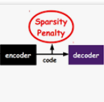
Point-of-Care Ultrasound (POCUS) refers to clinician-performed and interpreted ultrasonography at the patient's bedside. Interpreting these images requires a high level of expertise, which may not be available during emergencies. In this paper, we support POCUS by developing classifiers that can aid medical professionals by diagnosing whether or not a patient has pneumothorax. We decomposed the task into multiple steps, using YOLOv4 to extract relevant regions of the video and a 3D sparse coding model to represent video features. Given the difficulty in acquiring positive training videos, we trained a small-data classifier with a maximum of 15 positive and 32 negative examples. To counteract this limitation, we leveraged subject matter expert (SME) knowledge to limit the hypothesis space, thus reducing the cost of data collection. We present results using two lung ultrasound datasets and demonstrate that our model is capable of achieving performance on par with SMEs in pneumothorax identification. We then developed an iOS application that runs our full system in less than 4 seconds on an iPad Pro, and less than 8 seconds on an iPhone 13 Pro, labeling key regions in the lung sonogram to provide interpretable diagnoses.
相關內容
Learning diverse skills is one of the main challenges in robotics. To this end, imitation learning approaches have achieved impressive results. These methods require explicitly labeled datasets or assume consistent skill execution to enable learning and active control of individual behaviors, which limits their applicability. In this work, we propose a cooperative adversarial method for obtaining single versatile policies with controllable skill sets from unlabeled datasets containing diverse state transition patterns by maximizing their discriminability. Moreover, we show that by utilizing unsupervised skill discovery in the generative adversarial imitation learning framework, novel and useful skills emerge with successful task fulfillment. Finally, the obtained versatile policies are tested on an agile quadruped robot called Solo 8 and present faithful replications of diverse skills encoded in the demonstrations.
Even though auto-encoders (AEs) have the desirable property of learning compact representations without labels and have been widely applied to out-of-distribution (OoD) detection, they are generally still poorly understood and are used incorrectly in detecting outliers where the normal and abnormal distributions are strongly overlapping. In general, the learned manifold is assumed to contain key information that is only important for describing samples within the training distribution, and that the reconstruction of outliers leads to high residual errors. However, recent work suggests that AEs are likely to be even better at reconstructing some types of OoD samples. In this work, we challenge this assumption and investigate what auto-encoders actually learn when they are posed to solve two different tasks. First, we propose two metrics based on the Fr\'echet inception distance (FID) and confidence scores of a trained classifier to assess whether AEs can learn the training distribution and reliably recognize samples from other domains. Second, we investigate whether AEs are able to synthesize normal images from samples with abnormal regions, on a more challenging lung pathology detection task. We have found that state-of-the-art (SOTA) AEs are either unable to constrain the latent manifold and allow reconstruction of abnormal patterns, or they are failing to accurately restore the inputs from their latent distribution, resulting in blurred or misaligned reconstructions. We propose novel deformable auto-encoders (MorphAEus) to learn perceptually aware global image priors and locally adapt their morphometry based on estimated dense deformation fields. We demonstrate superior performance over unsupervised methods in detecting OoD and pathology.
Within medical imaging segmentation, the Dice coefficient and Hausdorff-based metrics are standard measures of success for deep learning models. However, modern loss functions for medical image segmentation often only consider the Dice coefficient or similar region-based metrics during training. As a result, segmentation architectures trained over such loss functions run the risk of achieving high accuracy for the Dice coefficient but low accuracy for Hausdorff-based metrics. Low accuracy on Hausdorff-based metrics can be problematic for applications such as tumor segmentation, where such benchmarks are crucial. For example, high Dice scores accompanied by significant Hausdorff errors could indicate that the predictions fail to detect small tumors. We propose the Weighted Normalized Boundary Loss, a novel loss function to minimize Hausdorff-based metrics with more desirable numerical properties than current methods and with weighting terms for class imbalance. Our loss function outperforms other losses when tested on the BraTS dataset using a standard 3D U-Net and the state-of-the-art nnUNet architectures. These results suggest we can improve segmentation accuracy with our novel loss function.
Image and text retrieval is one of the foundational tasks in the vision and language domain with multiple real-world applications. State-of-the-art approaches, e.g. CLIP, ALIGN, represent images and texts as dense embeddings and calculate the similarity in the dense embedding space as the matching score. On the other hand, sparse semantic features like bag-of-words models are more interpretable, but believed to suffer from inferior accuracy than dense representations. In this work, we show that it is possible to build a sparse semantic representation that is as powerful as, or even better than, dense presentations. We extend the CLIP model and build a sparse text and image representation (STAIR), where the image and text are mapped to a sparse token space. Each token in the space is a (sub-)word in the vocabulary, which is not only interpretable but also easy to integrate with existing information retrieval systems. STAIR model significantly outperforms a CLIP model with +$4.9\%$ and +$4.3\%$ absolute Recall@1 improvement on COCO-5k text$\rightarrow$image and image$\rightarrow$text retrieval respectively. It also achieved better performance on both of ImageNet zero-shot and linear probing compared to CLIP.
The automated detection of cancerous tumors has attracted interest mainly during the last decade, due to the necessity of early and efficient diagnosis that will lead to the most effective possible treatment of the impending risk. Several machine learning and artificial intelligence methodologies has been employed aiming to provide trustworthy helping tools that will contribute efficiently to this attempt. In this article, we present a low-complexity convolutional neural network architecture for tumor classification enhanced by a robust image augmentation methodology. The effectiveness of the presented deep learning model has been investigated based on 3 datasets containing brain, kidney and lung images, showing remarkable diagnostic efficiency with classification accuracies of 99.33%, 100% and 99.7% for the 3 datasets respectively. The impact of the augmentation preprocessing step has also been extensively examined using 4 evaluation measures. The proposed low-complexity scheme, in contrast to other models in the literature, renders our model quite robust to cases of overfitting that typically accompany small datasets frequently encountered in medical classification challenges. Finally, the model can be easily re-trained in case additional volume images are included, as its simplistic architecture does not impose a significant computational burden.
For medical image segmentation, contrastive learning is the dominant practice to improve the quality of visual representations by contrasting semantically similar and dissimilar pairs of samples. This is enabled by the observation that without accessing ground truth label, negative examples with truly dissimilar anatomical features, if sampled, can significantly improve the performance. In reality, however, these samples may come from similar anatomical features and the models may struggle to distinguish the minority tail-class samples, making the tail classes more prone to misclassification, both of which typically lead to model collapse. In this paper, we propose ARCO, a semi-supervised contrastive learning (CL) framework with stratified group sampling theory in medical image segmentation. In particular, we first propose building ARCO through the concept of variance-reduced estimation, and show that certain variance-reduction techniques are particularly beneficial in medical image segmentation tasks with extremely limited labels. Furthermore, we theoretically prove these sampling techniques are universal in variance reduction. Finally, we experimentally validate our approaches on three benchmark datasets with different label settings, and our methods consistently outperform state-of-the-art semi- and fully-supervised methods. Additionally, we augment the CL frameworks with these sampling techniques and demonstrate significant gains over previous methods. We believe our work is an important step towards semi-supervised medical image segmentation by quantifying the limitation of current self-supervision objectives for accomplishing medical image analysis tasks.
Object detectors usually achieve promising results with the supervision of complete instance annotations. However, their performance is far from satisfactory with sparse instance annotations. Most existing methods for sparsely annotated object detection either re-weight the loss of hard negative samples or convert the unlabeled instances into ignored regions to reduce the interference of false negatives. We argue that these strategies are insufficient since they can at most alleviate the negative effect caused by missing annotations. In this paper, we propose a simple but effective mechanism, called Co-mining, for sparsely annotated object detection. In our Co-mining, two branches of a Siamese network predict the pseudo-label sets for each other. To enhance multi-view learning and better mine unlabeled instances, the original image and corresponding augmented image are used as the inputs of two branches of the Siamese network, respectively. Co-mining can serve as a general training mechanism applied to most of modern object detectors. Experiments are performed on MS COCO dataset with three different sparsely annotated settings using two typical frameworks: anchor-based detector RetinaNet and anchor-free detector FCOS. Experimental results show that our Co-mining with RetinaNet achieves 1.4%~2.1% improvements compared with different baselines and surpasses existing methods under the same sparsely annotated setting.
A key requirement for the success of supervised deep learning is a large labeled dataset - a condition that is difficult to meet in medical image analysis. Self-supervised learning (SSL) can help in this regard by providing a strategy to pre-train a neural network with unlabeled data, followed by fine-tuning for a downstream task with limited annotations. Contrastive learning, a particular variant of SSL, is a powerful technique for learning image-level representations. In this work, we propose strategies for extending the contrastive learning framework for segmentation of volumetric medical images in the semi-supervised setting with limited annotations, by leveraging domain-specific and problem-specific cues. Specifically, we propose (1) novel contrasting strategies that leverage structural similarity across volumetric medical images (domain-specific cue) and (2) a local version of the contrastive loss to learn distinctive representations of local regions that are useful for per-pixel segmentation (problem-specific cue). We carry out an extensive evaluation on three Magnetic Resonance Imaging (MRI) datasets. In the limited annotation setting, the proposed method yields substantial improvements compared to other self-supervision and semi-supervised learning techniques. When combined with a simple data augmentation technique, the proposed method reaches within 8% of benchmark performance using only two labeled MRI volumes for training, corresponding to only 4% (for ACDC) of the training data used to train the benchmark.
Clustering is one of the most fundamental and wide-spread techniques in exploratory data analysis. Yet, the basic approach to clustering has not really changed: a practitioner hand-picks a task-specific clustering loss to optimize and fit the given data to reveal the underlying cluster structure. Some types of losses---such as k-means, or its non-linear version: kernelized k-means (centroid based), and DBSCAN (density based)---are popular choices due to their good empirical performance on a range of applications. Although every so often the clustering output using these standard losses fails to reveal the underlying structure, and the practitioner has to custom-design their own variation. In this work we take an intrinsically different approach to clustering: rather than fitting a dataset to a specific clustering loss, we train a recurrent model that learns how to cluster. The model uses as training pairs examples of datasets (as input) and its corresponding cluster identities (as output). By providing multiple types of training datasets as inputs, our model has the ability to generalize well on unseen datasets (new clustering tasks). Our experiments reveal that by training on simple synthetically generated datasets or on existing real datasets, we can achieve better clustering performance on unseen real-world datasets when compared with standard benchmark clustering techniques. Our meta clustering model works well even for small datasets where the usual deep learning models tend to perform worse.
Deep neural network architectures have traditionally been designed and explored with human expertise in a long-lasting trial-and-error process. This process requires huge amount of time, expertise, and resources. To address this tedious problem, we propose a novel algorithm to optimally find hyperparameters of a deep network architecture automatically. We specifically focus on designing neural architectures for medical image segmentation task. Our proposed method is based on a policy gradient reinforcement learning for which the reward function is assigned a segmentation evaluation utility (i.e., dice index). We show the efficacy of the proposed method with its low computational cost in comparison with the state-of-the-art medical image segmentation networks. We also present a new architecture design, a densely connected encoder-decoder CNN, as a strong baseline architecture to apply the proposed hyperparameter search algorithm. We apply the proposed algorithm to each layer of the baseline architectures. As an application, we train the proposed system on cine cardiac MR images from Automated Cardiac Diagnosis Challenge (ACDC) MICCAI 2017. Starting from a baseline segmentation architecture, the resulting network architecture obtains the state-of-the-art results in accuracy without performing any trial-and-error based architecture design approaches or close supervision of the hyperparameters changes.


