

In the last decade, convolutional neural networks (ConvNets) have been a major focus of research in medical image analysis. However, the performances of ConvNets may be limited by a lack of explicit consideration of the long-range spatial relationships in an image. Recently Vision Transformer architectures have been proposed to address the shortcomings of ConvNets and have produced state-of-the-art performances in many medical imaging applications. Transformers may be a strong candidate for image registration because their unlimited receptive field enables a more precise comprehension of the spatial correspondence between moving and fixed images. Here, we present TransMorph, a hybrid Transformer-ConvNet model for volumetric medical image registration. This paper also presents diffeomorphic and Bayesian variants of TransMorph: the diffeomorphic variants ensure the topology-preserving deformations, and the Bayesian variant produces a well-calibrated registration uncertainty estimate. We extensively validated the proposed models using 3D medical images from three applications: inter-patient and atlas-to-patient brain MRI registration and phantom-to-CT registration. The proposed models are evaluated in comparison to a variety of existing registration methods and Transformer architectures. Qualitative and quantitative results demonstrate that the proposed Transformer-based model leads to a substantial performance improvement over the baseline methods, confirming the effectiveness of Transformers for medical image registration.
相關內容
Removing noise from the any processed images is very important. Noise should be removed in such a way that important information of image should be preserved. A decisionbased nonlinear algorithm for elimination of band lines, drop lines, mark, band lost and impulses in images is presented in this paper. The algorithm performs two simultaneous operations, namely, detection of corrupted pixels and evaluation of new pixels for replacing the corrupted pixels. Removal of these artifacts is achieved without damaging edges and details. However, the restricted window size renders median operation less effective whenever noise is excessive in that case the proposed algorithm automatically switches to mean filtering. The performance of the algorithm is analyzed in terms of Mean Square Error [MSE], Peak-Signal-to-Noise Ratio [PSNR], Signal-to-Noise Ratio Improved [SNRI], Percentage Of Noise Attenuated [PONA], and Percentage Of Spoiled Pixels [POSP]. This is compared with standard algorithms already in use and improved performance of the proposed algorithm is presented. The advantage of the proposed algorithm is that a single algorithm can replace several independent algorithms which are required for removal of different artifacts.
Knowledge distillation (KD) has been actively studied for image classification tasks in deep learning, aiming to improve the performance of a student based on the knowledge from a teacher. However, applying KD in image regression with a scalar response variable has been rarely studied, and there exists no KD method applicable to both classification and regression tasks yet. Moreover, existing KD methods often require a practitioner to carefully select or adjust the teacher and student architectures, making these methods less flexible in practice. To address the above problems in a unified way, we propose a comprehensive KD framework based on cGANs, termed cGAN-KD. Fundamentally different from existing KD methods, cGAN-KD distills and transfers knowledge from a teacher model to a student model via cGAN-generated samples. This novel mechanism makes cGAN-KD suitable for both classification and regression tasks, compatible with other KD methods, and insensitive to the teacher and student architectures. An error bound for a student model trained in the cGAN-KD framework is derived in this work, providing a theory for why cGAN-KD is effective as well as guiding the practical implementation of cGAN-KD. Extensive experiments on CIFAR-100 and ImageNet-100 show that we can combine state of the art KD methods with the cGAN-KD framework to yield a new state of the art. Moreover, experiments on Steering Angle and UTKFace demonstrate the effectiveness of cGAN-KD in image regression tasks, where existing KD methods are inapplicable.
Remote-sensing (RS) Change Detection (CD) aims to detect "changes of interest" from co-registered bi-temporal images. The performance of existing deep supervised CD methods is attributed to the large amounts of annotated data used to train the networks. However, annotating large amounts of remote sensing images is labor-intensive and expensive, particularly with bi-temporal images, as it requires pixel-wise comparisons by a human expert. On the other hand, we often have access to unlimited unlabeled multi-temporal RS imagery thanks to ever-increasing earth observation programs. In this paper, we propose a simple yet effective way to leverage the information from unlabeled bi-temporal images to improve the performance of CD approaches. More specifically, we propose a semi-supervised CD model in which we formulate an unsupervised CD loss in addition to the supervised Cross-Entropy (CE) loss by constraining the output change probability map of a given unlabeled bi-temporal image pair to be consistent under the small random perturbations applied on the deep feature difference map that is obtained by subtracting their latent feature representations. Experiments conducted on two publicly available CD datasets show that the proposed semi-supervised CD method can reach closer to the performance of supervised CD even with access to as little as 10% of the annotated training data. Code available at //github.com/wgcban/SemiCD
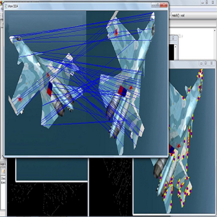
Cross-slide image analysis provides additional information by analysing the expression of different biomarkers as compared to a single slide analysis. These biomarker stained slides are analysed side by side, revealing unknown relations between them. During the slide preparation, a tissue section may be placed at an arbitrary orientation as compared to other sections of the same tissue block. The problem is compounded by the fact that tissue contents are likely to change from one section to the next and there may be unique artefacts on some of the slides. This makes registration of each section to a reference section of the same tissue block an important pre-requisite task before any cross-slide analysis. We propose a deep feature based registration (DFBR) method which utilises data-driven features to estimate the rigid transformation. We adopted a multi-stage strategy for improving the quality of registration. We also developed a visualisation tool to view registered pairs of WSIs at different magnifications. With the help of this tool, one can apply a transformation on the fly without the need to generate transformed source WSI in a pyramidal form. We compared the performance of data-driven features with that of hand-crafted features on the COMET dataset. Our approach can align the images with low registration errors. Generally, the success of non-rigid registration is dependent on the quality of rigid registration. To evaluate the efficacy of the DFBR method, the first two steps of the ANHIR winner's framework are replaced with our DFBR to register challenge provided image pairs. The modified framework produces comparable results to that of challenge winning team.
Biomedical Question Answering (BQA) has attracted increasing attention in recent years due to its promising application prospect. It is a challenging task because the biomedical questions are professional and usually vary widely. Existing question answering methods answer all questions with a homogeneous model, leading to various types of questions competing for the shared parameters, which will confuse the model decision for each single type of questions. In this paper, in order to alleviate the parameter competition problem, we propose a Mixture-of-Expert (MoE) based question answering method called MoEBQA that decouples the computation for different types of questions by sparse routing. To be specific, we split a pretrained Transformer model into bottom and top blocks. The bottom blocks are shared by all the examples, aiming to capture the general features. The top blocks are extended to an MoE version that consists of a series of independent experts, where each example is assigned to a few experts according to its underlying question type. MoEBQA automatically learns the routing strategy in an end-to-end manner so that each expert tends to deal with the question types it is expert in. We evaluate MoEBQA on three BQA datasets constructed based on real examinations. The results show that our MoE extension significantly boosts the performance of question answering models and achieves new state-of-the-art performance. In addition, we elaborately analyze our MoE modules to reveal how MoEBQA works and find that it can automatically group the questions into human-readable clusters.
Transformers have dominated the field of natural language processing, and recently impacted the computer vision area. In the field of medical image analysis, Transformers have also been successfully applied to full-stack clinical applications, including image synthesis/reconstruction, registration, segmentation, detection, and diagnosis. Our paper presents both a position paper and a primer, promoting awareness and application of Transformers in the field of medical image analysis. Specifically, we first overview the core concepts of the attention mechanism built into Transformers and other basic components. Second, we give a new taxonomy of various Transformer architectures tailored for medical image applications and discuss their limitations. Within this review, we investigate key challenges revolving around the use of Transformers in different learning paradigms, improving the model efficiency, and their coupling with other techniques. We hope this review can give a comprehensive picture of Transformers to the readers in the field of medical image analysis.
Applying artificial intelligence techniques in medical imaging is one of the most promising areas in medicine. However, most of the recent success in this area highly relies on large amounts of carefully annotated data, whereas annotating medical images is a costly process. In this paper, we propose a novel method, called FocalMix, which, to the best of our knowledge, is the first to leverage recent advances in semi-supervised learning (SSL) for 3D medical image detection. We conducted extensive experiments on two widely used datasets for lung nodule detection, LUNA16 and NLST. Results show that our proposed SSL methods can achieve a substantial improvement of up to 17.3% over state-of-the-art supervised learning approaches with 400 unlabeled CT scans.
The U-Net was presented in 2015. With its straight-forward and successful architecture it quickly evolved to a commonly used benchmark in medical image segmentation. The adaptation of the U-Net to novel problems, however, comprises several degrees of freedom regarding the exact architecture, preprocessing, training and inference. These choices are not independent of each other and substantially impact the overall performance. The present paper introduces the nnU-Net ('no-new-Net'), which refers to a robust and self-adapting framework on the basis of 2D and 3D vanilla U-Nets. We argue the strong case for taking away superfluous bells and whistles of many proposed network designs and instead focus on the remaining aspects that make out the performance and generalizability of a method. We evaluate the nnU-Net in the context of the Medical Segmentation Decathlon challenge, which measures segmentation performance in ten disciplines comprising distinct entities, image modalities, image geometries and dataset sizes, with no manual adjustments between datasets allowed. At the time of manuscript submission, nnU-Net achieves the highest mean dice scores across all classes and seven phase 1 tasks (except class 1 in BrainTumour) in the online leaderboard of the challenge.
In this paper, we adopt 3D Convolutional Neural Networks to segment volumetric medical images. Although deep neural networks have been proven to be very effective on many 2D vision tasks, it is still challenging to apply them to 3D tasks due to the limited amount of annotated 3D data and limited computational resources. We propose a novel 3D-based coarse-to-fine framework to effectively and efficiently tackle these challenges. The proposed 3D-based framework outperforms the 2D counterpart to a large margin since it can leverage the rich spatial infor- mation along all three axes. We conduct experiments on two datasets which include healthy and pathological pancreases respectively, and achieve the current state-of-the-art in terms of Dice-S{\o}rensen Coefficient (DSC). On the NIH pancreas segmentation dataset, we outperform the previous best by an average of over 2%, and the worst case is improved by 7% to reach almost 70%, which indicates the reliability of our framework in clinical applications.
Deep learning (DL) based semantic segmentation methods have been providing state-of-the-art performance in the last few years. More specifically, these techniques have been successfully applied to medical image classification, segmentation, and detection tasks. One deep learning technique, U-Net, has become one of the most popular for these applications. In this paper, we propose a Recurrent Convolutional Neural Network (RCNN) based on U-Net as well as a Recurrent Residual Convolutional Neural Network (RRCNN) based on U-Net models, which are named RU-Net and R2U-Net respectively. The proposed models utilize the power of U-Net, Residual Network, as well as RCNN. There are several advantages of these proposed architectures for segmentation tasks. First, a residual unit helps when training deep architecture. Second, feature accumulation with recurrent residual convolutional layers ensures better feature representation for segmentation tasks. Third, it allows us to design better U-Net architecture with same number of network parameters with better performance for medical image segmentation. The proposed models are tested on three benchmark datasets such as blood vessel segmentation in retina images, skin cancer segmentation, and lung lesion segmentation. The experimental results show superior performance on segmentation tasks compared to equivalent models including U-Net and residual U-Net (ResU-Net).
小貼士
登錄享
相關主題


注冊地址: 北京市海淀區羊坊店路18號2幢3層301-191